Slide #DMS 126 [Esophagus, Monkey, PTS]. This is from the upper 1/3 of the monkey esophagus. Diagnostic features are the stratified squamous lining and the presence of a muscularis mucosae. Look for scattered submucosal glands; these are characteristic of the esophagus but are not always cut in every section. Does your section have ducts? secretory portions? If present, are the glands mucous or serous? If a duct is present, check whether it has a two-layered stratified cuboidal epithelium. Compare slides with a neighbor.
What is the advantage of having stratified squamous epithelium line the lumen of the esophagus? Is it keratinized?
Notice that the muscularis externa is composed of both smooth and skeletal muscle. Typically, in the human, the upper third of the esophagus has wholly skeletal muscle, the lower third wholly smooth muscle, and the middle third a mixture of the two. Functionally, what does this mean?
As you see blood vessels in the connective tissue layers of the wall, practice identifying them as arteries or veins. Can you also find nerves and autonomic (parasympathetic) ganglia (hint: there are some in the submucosa and some at the junction between longitudinal and circular muscle)?
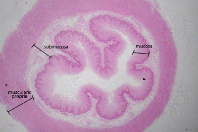
This is a low power view of a section through the esophagus. As with other parts of the GI tract, one can usually define the four layers of the gut wall, the mucosa, submucosa, muscularis propria (externa) and an outermost serosa or adventitial layer, which is not easily seen in this particular image.
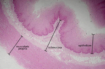
A higher power view of the esophagus reveals the non-keratinized, stratified squamous epithelium that makes up the bulk of the mucosal layer (a lamina propria and muscularis mucosa are not very obvious in this preparation). The submucosa is a connective tissue layer in which many blood vessels may be found. Note the extension of the submucosal tissue into the mucosa, creating macroscopic fold (plica) of the mucosal surface. External to the submucosa, one finds the muscle tissue of the muscularis propria. Depending on where along the length of the esophagus this section was taken, this muscle may be striated, smooth, or a combination of the two.
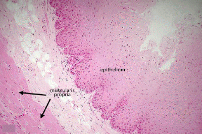
At higher power, one can better define the stratified squamous epithelium of the esophagus. Distal to the esophagus, the epithelium lining the GI tract will be a simple columnar one until the stratified squamous type returns at the level of the anus. In this view, it is apparent that the muscularis propria consists of skeletal muscle, as peripherally-located nuclei are apparent. Thus, this section of esophagus was taken from its more proximal portion.