Slide #DMS 127 [Human esophagus - mid portion]. This is from the mid-portion of the human esophagus. Diagnostic features are the stratified squamous lining and the presence of a muscularis mucosae. Look for scattered submucosal glands; these are characteristic of the esophagus but are not always cut in every section. Does your section have ducts? secretory portions? If present, are the glands mucous or serous? If a duct is present, check whether it has a two-layered stratified cuboidal epithelium. Compare slides with a neighbor.
What is the advantage of having stratified squamous epithelium line the lumen of the esophagus? Is it keratinized?
Notice that the muscularis externa is composed of both smooth and skeletal muscle. Typically, in the human, the upper third of the esophagus has wholly skeletal muscle, the lower third wholly smooth muscle, and the middle third a mixture of the two. Functionally, what does this mean?
As you see blood vessels in the connective tissue layers of the wall, practice identifying them as arteries or veins. Can you also find nerves and autonomic (parasympathetic) ganglia (hint: there are some in the submucosa and some at the junction between longitudinal and circular muscle)?
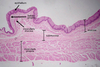
This is a low power image of a different section of esophagus. In this example, all three layers of the mucosa may be defined: the epithelium, the lamina propria, and the muscularis mucosa. External to the mucosa one will again find the submucosa and then the muscularis propria (muscularis externa). Again, the outermost adventitial layer is not easily appreciated in this section.
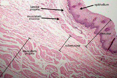
At slightly higher power, again define the three layers that make up the mucosa: the stratified squamous epithelium, the underlying loose connective tissue of the lamina propria, and then the smooth muscle of the muscularis mucosa. Submucosal tissue and the smooth muscle of the muscularis propria of this distal portion of the esophagus can be seen.