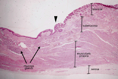
Slide #DMS 128 [Dog - Gastroesophageal junction, H&E]. This longitudinal section illustrates the transition between lower (subdiaphragmatic esophagus) and the so-called cardiac portion of the stomach. Again define the four basic layers. Note the rather abrupt transistion in type of lining epithelium. The so-called cardiac region of the stomach is lined by simple columnar epithelium that extends into the lamina propria forming the shallow, mucus and lysozyme-secreting cardiac glands. Just beyond, the deeper and more typical gastric glands are evident in the fundic portion of the stomach. Can you find the lamina propria and muscularis mucosa (smooth muscle cut longitudinally). In many of the sections, the submucosa of the lower esophagus has well developed mucous glands. Note also the large blood vessels in the submucosa at the junctional zone . Relationship to "esophageal varices"? Depending on your section, you may see a gradual transition from smooth to skeletal muscle in the muscularis externa, or the entire muscularis may be of smooth muscle composition. Is the outer layer of the esophagus and stomach serosal or adventitial?
The walls may be thrown into so many folds and cut so tangentially, that relationships are not immediately clear. However, if you will find the esophageal end and follow along the length of its luminal surface, you will find at some point a very sharp transition from stratified squamous to a simple columnar epithelium. Other coats of the wall have more of a zone of overlap and transition.
Look for submucosal glands in the esophagus and mucosal (cardiac) glands in the stomach; notice their positional relationship to the muscularis mucosae in each instance. In the muscularis externa is the muscle wholly smooth, or is it something skeletal? (Animals vary in this respect.)

This is a low power view of the junction between the esophagus on the left and the stomach on the right. The transition in the surface epithelium is marked by the arrowhead. Again, define the four layers of the gut wall, mucosa, submucosa, muscularis propria and adventitia/serosa. Note the submucosal mucous glands in this distal portion of the esophagus that terminate, roughly, where the stomach begins.
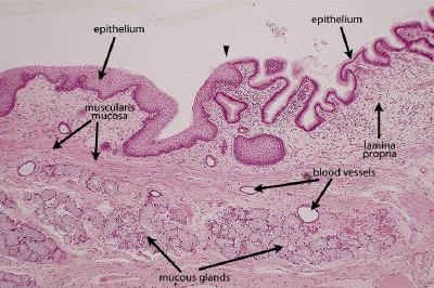
This is a medium power view of the mucosa and a portion of the submucosa at the gastroesophageal junction. The epithelial transition, from stratified squamous in the esophagus to simple columnar in the stomach, is indicated by the arrowhead. The three layers of the mucosa, epithelium, lamina propria and muscularis mucosa are all clearly seen. Note the submucosal mucous glands in the esophagus, and some of the numerous blood vessels in the submucosal layer.
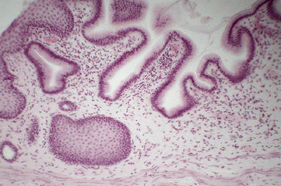
This is a view of the mucosal layer at the border between the esophagus and the stomach. Note the change in the type of epithelium lining the lumen of these two organs: stratified squamous in the esophagus and simple columnar in the stomach. The proximal most part of the stomach (the cardia) is characterized by mucous-secreting cardiac glands, seen in a different preparation in the next image.
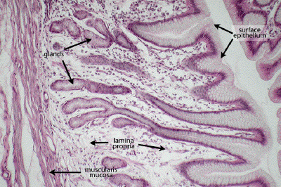
This is a medium power view through the cardiac portion of the stomach. Like all segments of the stomach, the surface epithelium consists of a homogenous population of surface mucous (foveolar) cells. In the cardiac portion of the stomach, the glands are also primarily mucus-producing as seen here. Note the loose connective tissue of the lamina propria and the smooth muscle of the muscularis mucosa.
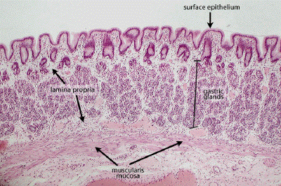
Moving from the cardia into the body (corpus) or fundic region of the stomach, note the replacement of the mucous-secreting cardiac glands with the more heterogeneous gastric (oxyntic) glands, the structure of which will be examined in more detail in subsequent images. The surface epithelium, however, still consists of mucus-secreting surface foveolar cells as it does throughout the entirety of the stomach. A bit of lamina propria fills the spaces between the gastric glands, and a well-defined muscularis mucosa separates the mucosa from the underlying submucosal tissue.