
Slide #DMS 129 [Stomach, Human, PTS]. Identify the mucosa, submucosa, muscularis externa and serosa. The surface mucous cells of the mucosa are well shown. How do they differ from mucous secreting goblet cells? Identify gastric pits and gastric glands. Define the lamina propria. The parts of gastric glands are isthmus, neck , and base. What kinds of cells are typically found in each of these parts. Where are the "regenerative cells" of the gastric epithelium located? How frequently are the surface epithelial cells replaced? In this section, chief cells and parietal cells can be distinguished from each other since the chief cells show a cytoplasmic basophilia. Why? Study the gastric glands. What do each of these cell types secrete? Review the E.M. characteristics of these cells.
Enteroendocrine cells are also present in the basal part of the glands, but are positively identified only with the use of appropriate immunochemical markers. Be aware of the many endocrine/paracrine factors these cells secrete. Can you find any ganglion cells of the myenteric (Auerbach's) plexus between the muscle layers?
Find an area of mucosa where you can see the whole length of the long, straight, tubular gastric glands. Note that the simple columnar mucin-secreting cells of the stomach surface continue down into the open pits (or foveolae) of the glands for just a short distance.
Where are the connective tissue fibers and cells of the lamina propria? Where is the muscularis mucosae in relation to the glands? Look for strands of smooth muscle passing up into the lamina propria, between the glands
Notice that the submucosa extends up into the temporary folds (or rugae) which protrude into the lumen of the stomach. Many blood vessels run in the submucosa.
In what direction do the muscle fibers of the muscularis externa run? Is the outermost layer an adventitia or serosa?
A diagnostic feature to remember: the stomach epithelium contains no goblet cells; that of the intestine does.

This is a very low power image of a plastic thin section taken from the body or fundic portion of the stomach. The four layers typical of the GI tract can be defined: the mucosa, submucosa, muscularis propria, and, in this case, a serosa. Even at this low power, one can see the variable staining pattern of the glandular epithelium of the stomach, reflecting the pattern of distribution of discrete cell types within this epithelium.
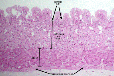
This is a low power view of the mucosal layer of the body or fundus of the stomach. The muscularis mucosa, separating the mucosa from the underlying submucosal tissue can be seen. Note the distinct staining pattern of the glandular epithelium, reflecting the different populations of cells found 1) at the surface and in the gastric pits (surface foveolar cells), 2) in the isthmus and neck portions of the glands (mostly mucous neck cells and parietal [oxyntic] cells), and 3) at the base of the glands (chief [zymogenic] cells). Note also the very sparse lamina propria. While all the cells of the gastric gland secrete into the same lumen, the continuity of the gland from base to surface is rarely appreciated in section.
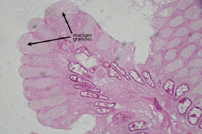
This is a high power view of the surface mucous (or foveolar) cells that one would find lining the surface and gastric pits of the stomach. Note the abundant mucigen granules found in the cytoplasm of all of these cells.
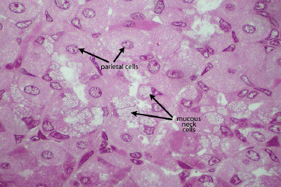
This is a medium power view of the cell population one would find in the isthmus and neck of a gastric (oxyntic) gland. Both mucous neck cells (an additional source of mucus for the stomach) and the large, eosinophilic parietal (oxyntic) cells can be seen. The parietal cells are the source of both hydrochloric acid (HCl) and gastric intrinsic factor. A population of stem cells would also be found in the neck of the gastric glands, but are difficult to appreciate in the absence of mitotic figures.
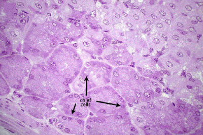
In this image, appreciate the change in cell type as one moves from the neck of the gland towards the base of the gastric gland. The base of the gland is dominated by the basophilic chief (zymogenic) cells, seen in more detail in the next image. Enteroendocrine cells would also be found in the base of the glands, but these cells are difficult to distinguish in the absence of specific immunochemical stains.
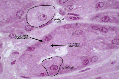
A high power view of the base of a gastric gland shows the chief (zymogenic) cells found in this location. Note the basophilic cytoplasm, reflective of an abundance of rough endoplasmic reticulum, and the numerous zymogen granules found in the apical cytoplasm. A parietal cell is also seen in this image for comparison. The mottling of the cytoplasm of the parietal cell reflects the intracellular canalicular system in that cell that greatly increases the surface area for the secretion of HCl by that cell type.