Slide #DMS 130 [Dog; gastroduodeneal junction, H&E]. This slide shows well the transition from pyloric region of stomach to duodenum (1st part). Important to scan this slide with your 4x objective first. Try to find the exact point of transition, marked by distinct changes in mucosa and other morphological features. Study first the pyloric region of stomach. Note that the gastric glands are different from those of the fundic/body part of the stomach. They are much deeper and contain mostly mucous secreting cells. The muscularis of the stomach is usually described as three-layered: inner oblique, middle circular, outer longitudinal. However, in sections the distinction is often difficult to make. Which layer appears to hypertrophy to form the pyloric sphincter?
Now look at the duodenal portion of the slide. Identify the layers. What feature is the most reliable for identifying the duodenum from other parts of the small bowel? The villi of the initial part of the duodenum are short, broad and rather atypical.
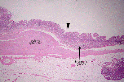
This is a very low power view of the junction between stomach (left) and duodenum (right). The transition occurs in the area of the arrowhead. Note the thickening of the inner circular layer of smooth muscle at the distal end of the stomach which constitutes the pyloric sphincter. Note also the appearance of submucosal glands (Brunner's glands) which are a hallmark of the duodenal portion of the small intestine.
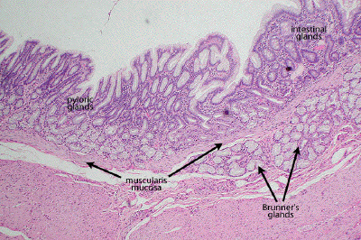
At low power, note the mucous secreting glands typical of the pyloric (antral) portion of the stomach. These glands will give way to the intestinal glands more typical of the duodenum and other portions of the intestinal tract. Note the well-defined muscularis mucosa, separating the mucosa of the gut from the submucosal tissues, including the submucosal Brunner's glands of the duodenum.
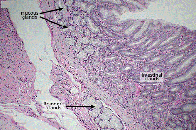
At this higher power, one may better appreciate the mucous secreting glands typical of the antral portion of the stomach and the development of intestinal glands as one enters the duodenum. Note the beginning of the submucosal Brunner's glands that typify the duodenum. These glands produce a bicarbonate-rich mucous which helps to neutralize the acidic stomach contents.
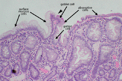
This is a medium power view that shows the transition (arrowhead) in the epithelium as one moves from the stomach (left) to the duodenum (right). Unlike the stomach, whose surface is covered by a homogenous population of surface mucous (foveolar) cells, the surface epithelium of the intestinal tract shows absorptive cells as well as some number of mucus-secreting goblet cells.