Slide #DMS 135 [Large intestine, monkey, mucus stain]. This section has been stained by the mucicarmine technique which stains mucus (and mucinogen) red. First of all note the complete lack of villi (typical of large bowel). Don't mistake intestinal glands for villi. The epithelium again consists of absorptive and goblet cells. The goblet cells are even more abundant than in the small intestine. Looking at the material in the lumen one can get a good idea of the functional importance of goblet cell mucus secretion. Lymphoid nodules are present in the submucosa. Can you see any taeniae coli?
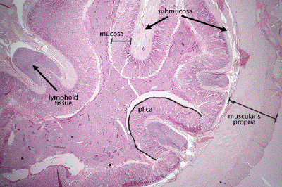
This is a very low power view through a section of colon (large intestine). Mucosa, submucosa and muscularis propria (externa) can be defined. Some lymphoid follicles are seen in the submucosal tissue. Note that both submucosa and mucosa are thrown into large, macroscopic folds or plicae. Villi are NOT a feature of the colon.
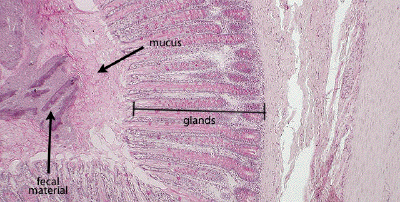
This is a low power view of the mucosa of the colon. Note that villi are not seen in the colon, only colonic glands (crypts), which are replete with goblet cells (stained pink). The abundance of mucus in the lumen (surrounding fecal material) is evidence of the secretory function of these glands.
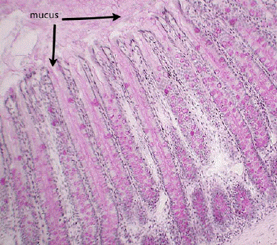
At medium power, note the abundance of goblet cells lining the glands of the colon. Their mucous component is stained pink in this preparation. One can almost watch the mucus pouring from the opening of the glands into the lumen of the colon.