Slide #DMS 137A [Human; appendix, H&E]. Like the large intestine, the appendix has no villi but does have crypts with many goblet cells. As seen here, the crypts are few in number and widely scattered in the mucosa. The main diagnostic feature of the appendix is the diffuse lymphatic tissue pervading the mucosa and submucosa, and sometimes including scattered nodules. Can you find any remnants of muscularis mucosae? Note that there are no taenia in the appendix. Note that the lumen of the appendix is filled with debris (including neutrophils, ?pus).
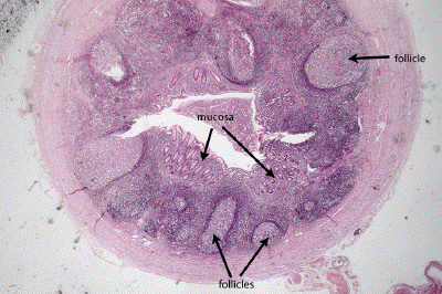
This is a very low power view of a section through the appendix. Note the great abundance of lymphoid tissue in the submucosa; several secondary lymphoid follicles with germinal centers can be seen. A typical colonic mucosa may be seen as well.
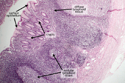
At somewhat higher power, again note the abundance of lymphoid tissue, both diffuse and nodular, present throughout the mucosa and submucosa of the appendix. Some of the surface epithelium and crypts may be seen. The mucosa of the appendix is comparable to that of the rest of the colon in that it lacks villi.
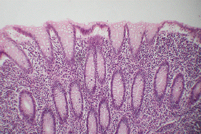
At medium power, note the typical colonic epithelium covering the surface and forming the glands (crypts) seen here. This is a simple columnar epithelium replete with goblet cells. Appreciate the abundant, diffuse lymphatic tissue infiltrating the lamina propria of the mucosa and surrounding the crypts.