Slide #DMS 148 [Gallbladder, Monkey, H&E+OG]. Observe that the mucosa consists of a simple columnar epithelium and a lamina propria. The lamina propria here was studied earlier as a well preserved example of loose (areolar) connective tissue. There is no muscularis mucosa or submucosa. Note that the muscularis is in irregular bundles that does not show the inner circular and outer longitudinal arrangement found in the gut. External to the muscularis is some moderately dense connective tissue. Using higher power, return to the mucosa. Note that it is thrown up into folds. The columnar epithelial cells are quite tall. Goblet cells are not normally found in the epithelium. A section of gall bladder should not be confused with sections taken elsewhere in the gastro-intestinal tract. The gall bladder has no villi, no crypts, no muscularis mucosa and no goblet cells in its epithelium. There is an external serosa with a mesothelial surface. What is the function of the gall bladder?
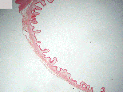
This slide presents a very low power view of a section through monkey gall bladder stained with H&E plus Orange G. Note that the mucosal surface is thrown into a series of folds or ridges which have a bit of similarity to the villus architecture of the small bowel. These structures, however, are NOT villi.
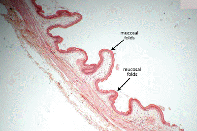
At low power, observe the irregular mucosal folds (NOT villi, there are NO crypts) and the poorly organized layers making up the remainder of the wall of the gall bladder.
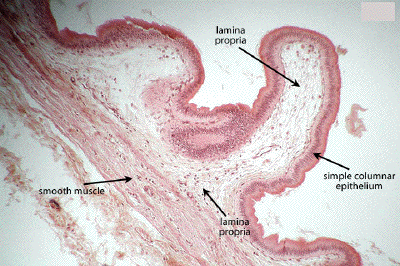
At medium power, define the simple columnar epithelium covering the mucosal folds. With the absence of a muscularis mucosa, there is no submucosa, only a lamina propria. The smooth muscle in the wall of the gall bladder is poorly organized and does not arrange itself into the more well-defined muscular layers seen in other GI organs. The outermost layer of the gall bladder may be covered by an adventitia (if in contact with the liver) or by a serosa (the peritoneal surface).
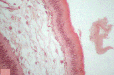
At high power, note the homogeneity of the simple columnar epithelium of the gall bladder. Goblet cells are NOT seen in the epithelium and help to distinguish this organ from the small intestine with which it might sometimes be confused.