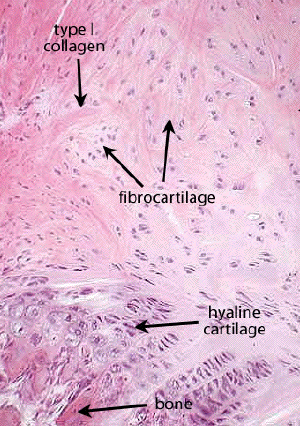
Slide DMS066 [Symphysis pubis, young rabbit; H&E]. The tissue at the periphery is mostly mature skeletal muscle. Developing bone is present in triangular wedges on each side of the midline cartilaginous symphysis.
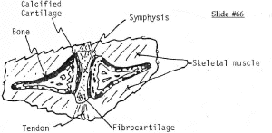
In most sections the junction of developing bone is marked by a prominent basophilic zone of calcified cartilage. The muscle attaches to the periosteum of the bone and the symphysis be means of tendinous insertions. As indicated in the diagram, the symphysis is a cartilaginous joint (synchondrosis) of limited mobility (amphiarthrosis). (Same as an intervertebral disc). The center of the joint is mostly hyaline cartilage. There is a transition to fibrocartilage as the inferior and superior margins of the symphysis and this in turn makes a transition to dense fibrous CT (tendon).
Under hormonal influence the cartilage of the pubic symphysis becomes markedly more flexible during the late stages of pregnancy. The symphysis may be completely calcified or ossified in late adulthood.