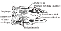
Slide DMS065 [Larynx]. This is a frontal section of the larynx of a cat or rabbit. (May include part of thyroid gland). Orient yourself to this section using low power and by referring to the diagram below.
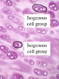
Find the areas of mature hyaline cartilage which compose the largest laryngeal plates and the tracheal rings. Identify matrix, chondrocytes within lacunae, and note the occurrence of cell nests. Cell nests result from the mitotic division of chondrocytes and are a manifestation of interstitial growth.
Chondrocytes are usually shrunken and distorted because a rather long time is required for the fixative to penetrate the dense matrix. Note the deep basophilia of the cartilage matrix immediately surrounding the chondrocytes. This constitutes the territorial matrix (capsular matrix), as distinguished from the more lightly stained interterritorial matrix found more peripherally. What is the explanation for the different staining properties of the cartilage matrix?
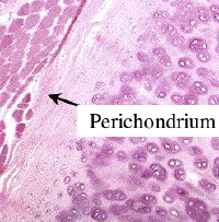
Delineate the perichondrium. Note that the chondrogenic layer is less prominent than in immature hyaline cartilage. Observe that capillaries are not present in the matrix of cartilage. Thus, chondrocytes must be nourished by slow diffusion from blood vessels located in the fibrous layer of the perichondrium. Cartilage has a low rate of metabolic activity and, following injury, regeneration may be very slow or absent. Note areas of calcification within the central regions of some of the pieces of cartilage.
Cartilage, unlike bone, can enlarge by interstitial growth as well as appositional growth. Where do these two growth processes occur?
One may be able to find an example of elastic cartilage stained only with H&E on the left side of this slide (in the epiglottal cartilage).