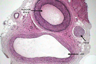
Slide #DMS 108 [Medium-size, muscular artery, Weigert stain]. This slide contains a complete neurovascular bundle (artery, vein and nerve). First, examine the medium-size, distributing ("muscular") artery. In general, the muscular arteries correspond to the smaller named arteries which you dissected in gross anatomy lab. Contrast in detail the structure of the media of these arteries to that of the aorta. Note the predominance of smooth muscle, the sparseness of elastic fibers and the prominent internal elastic lamina. What is the significance of the difference in relative amounts and organization of elastic fibers, collagenous fibers, and smooth muscle cells in terms of the functional role played by the two types of arteries in controlling circulation?
Note that the adentitia of these muscular arteries contains small blood vessels (vasa vasorum) and elastic and collagen fibers.

This very low power view of a neurovascular bundle shows a medium-sized muscular artery, its accompanying vein, and a small nerve. The Verhoeff's stain highlights the elastic fiber components (darkly stained).

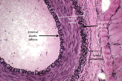
This low power view of a portion of a muscular artery, stained for elastic tissue, highlights the characteristic structure of this class of blood vessel: a prominent internal elastic lamina marking the outer limit of the tunica intima, a tunica media made up of many layers of circularly-arranged smooth muscle cells, a somewhat ill-defined external elastic lamina marking the outer border of the media, and the tunica adventitia which blends in with the perivascular connective tissue.

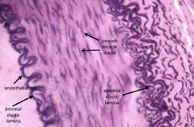
In this image of the inner walls of a muscular artery, the elastic stain highlights the characteristic scalloped appearance of the internal elastic lamina (IEL). This scalloping is an artifact of the agonal contraction of the blood vessel at the time of death/fixation. One can make out a few endothelial cell nuclei covering the luminal surface of the IEL. The cork-screw shaped nuclei of the smooth muscle cells dominating the tunica intima is apparent, as are the redundant layers of the external elastic lamina, marking the outer border of the media.
Next, try to identify veins. Study the relative thickness of the tunics in the walls of these veins and compare their characteristics with those of the comparable muscular artery as observed above. Next compare the distribution of elastic fibers. As compared with the accompanying artery, how do you relate the larger caliber and thinner wall of the vein to its function? Veins vary greatly in their histology according to the amount of hydrostatic pressure exerted on the wall. Thus, those located above the heart have a thinner wall than those located below the heart.

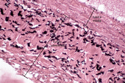
In this image of a medium-sized vein, stained for elastic tissue, one should again notice that the tunica adventitia makes up the bulk of the wall, not the tunica media as would be the case in a comparably-sized artery. Bundles of elastic fibers are seen in the adventitia, and, like other elements in this tunic, are longitudinally disposed (i.e. along the long axis of the blood vessel).