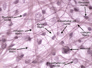
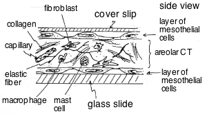
Slide DMS53 [Mesentary whole mount]. Loose CT (aka areolar ["small space"] CT). Study Slide #53 of a whole piece (not a section) of mesentery stained with resorcin-fuchsin. The mesentery (which attaches the small intestines to the body wall) is covered on both sides by a layer of mesothelial cells, but only the nuclei of these cells are usually seen. They are the largest nuclei present; their chromatin is seen as very fine granules, and usually a single nucleolus is visible. Between the mesothelial layers on both sides of the piece of tissue is loose, irregularly arranged (areolar) connective tissue (see diagram below). Many fibroblasts and perhaps a few macrophages are present. Fibroblasts have oval nuclei that are larger and have finer chromatin material than do the nuclei of macrophages. Often surrounding the dense, irregular nuclei of the macrophages are dark particles which the macrophages have phagocytosed and which lie in their cytoplasm. Do not spend an inordinate amount of time trying to distinguish macrophages in this preparation as you will see much better examples in subsequent slides. A number of mast cells are present. Their cytoplasm is usually so filled with metachromatic (purplish) granules that their rather small, round nuclei are often not visible. (Define the term metachromasia.) A small number of other cell types (e.g., white blood cells) not pertinent to this study are present also in this connective tissue. Collagen fibers (Type I) are visible as thick, acidophilic (pinkish-purple) strands extending throughout the tissue. The much thinner, dark purple-stained, branching strands are elastic fibers. (Blood capillaries, venules or arterioles may course through the connective tissue in some of the slides.)
Also look for the stretched thin, branching, dark-purple stained elastic fibers in this tissue.