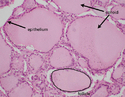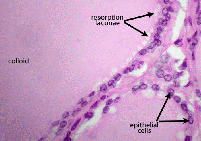
Slide #DMS 159 [Thyroid, human, H&E]. Thyroid parenchymal cells characteristically are arranged in a cyst-like fashion around a central storage vacuole usually filled with a highly eosinophilic secretory product called colloid. (The parallel cracks in the colloid are preparation artifacts). The colloid consists mainly of thyroglobulin, the storage form of the hormones T3 and T4. The cyst-like bodies are called follicles. Study the follicles in detail. The size of the follicles varies with their physiological activity as well as with the plane of sectioning. Note the more or less simple cuboidal follicular epithelium. The height of the epithelium also varies with physiological activity; in general, the higher the epithelium the greater the activity. Look for ìactive folliclesî showing ìreabsorption lacunaeî.
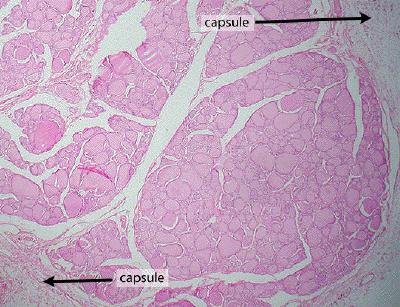
The thyroid has a relatively delicate capsule of fibroelastic connective tissue which penetrates between the follicles to form the stroma of the gland. Typical of endocrine glands, the stroma is well vascularized.

This is a very low power view of a section through the thyroid gland. Note the loose connective tissue capsule, trabeculae of which organize the parenchyma into rather irregular lobules.

At medium power, one observes the thyroid follicles that form the bulk of the gland. The follicles are filled with an eosinophilic colloid, the predominant component of which is thyroglobulin. The follicle is defined by the follicular epithelium which may vary in height depending on the activity state of the follicle. Numerous blood vessels course throughout the connective tissue between the follicles.

A high power view through portions of three thyroid follicles demonstrates the follicular epithelial cells, the eosinophilic colloid material, as well as resorption lacunae resulting from the uptake of colloid by the follicular epithelial cells. Parafollicular cells are also a component of the follicular wall, but cannot be distinguished (except in dogs) without the use of specific immunocytochemical markers.