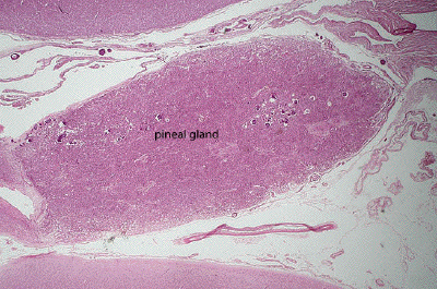
Slide #DMS 166 [Pineal gland]. The pineal gland is an organ that is comprised of modified retinal cells (pinealocytes) and glial cells. The pinealocytes release melatonin into the circulation in a circadian fashion, under the control of the sympathetic nervous system (in lower species they directly respond to light).
This is a very low power view of a section through the pineal gland and surrounding brain tissue. The pineal is located in the midline of the brain near the posterior wall of the third ventricle (just to the left of this picture).
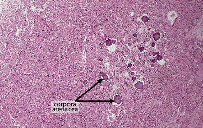
A low power view of the pineal reveals its most distinctive feature, the calcified concretions known as corpora arenacea or, more affectionately, brain sand. These concretions are radio-opaque and thus may appear in radiographs of the brain, particularly in older people.
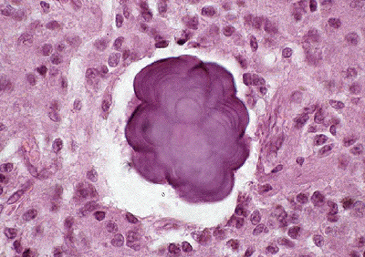
A high power view of the pineal shows a concretion and the surrounding parenchymal cells of the pineal gland, known as pinealocytes, together with their supporting glial cells.