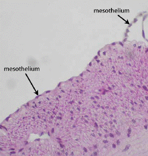
Slide DMS037. Look at this slide of the G.I. tract [small intestine, hamster; P.A.S. + H] with high magnification. Observe the abluminal (non-villous) surface and note a delicate, thin layer of simple squamous epithelial cells (may be artefactually missing in some places). Generally, only the flattened nuclei of this layer will be seen; the cytoplasm is too attenuated to be seen in section. This layer of simple squamous epithelium is part of the visceral peritoneum (mesothelium) that covers the surface of many of the organs within the peritoneal cavity.
Now observe the luminal surface. This consists of simple columnar epithelium with goblet cells. The goblet cells are examples of unicellular (single cell) glands.
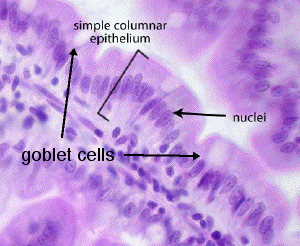
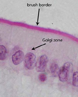
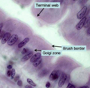
The surface of the simple columnar cells is covered with microvilli. The brush borders of these cells are distinctly "P.A.S. positive" (red staining). Why? The actin filament cores of the microvilli extend down into the apical cytoplasm of the cell where they are crosslinked by myosin and other actin- associated proteins. This dense meshwork of filaments is the terminal web which can be see as the densely eosinophilic line immediately beneath the microvilli.
The H&E picture (below) shows the specializations of the coumnar cells at very high power. Note the goblet cell on the right.
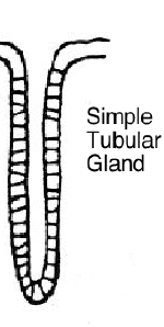
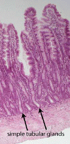
Examples of simple tubular glands may be seen in this section of the small intestine. Locate longitudinal and cross sections of some of these glands (located subjacent to villi). Most of the cells lining each simple gland are goblet cells.