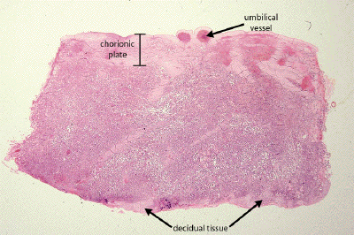
Slide DMS183 [Placenta, term, human, H&E]. This is an excellent section of a delivered term placenta and worth careful study. Once more identify the chorionic plate (better named "chorioamnionic plate" since it consists of fused chorion and amniotic membrane). An intact aminiotic membrane covers the fetal surface (it is continuous with amniotic covering of the umbilical cord that attaches to the the chorionic plate). At the fetal suface find the origin of stem villi and, extending into the intervillous space, their branches: terminal villi and anchoring villi. Note that the outer covering of the villi is at this stage almost exclusively syncytiotrophoblast. In the smallest terminal villi note the proximity of fetal capillaries to the syncytiotrophoblast. Of what would the minimal hemochorial placental barrier consist?
At the side opposite the chorionic plate in this section there remains attached some decidua basalis (maternal tissue) consisting of large decidual cells and abundant eosinophilic fibrin material (so-called Nitabuch's layer). Note the attachment of anchoring villi to this layer. Where does detachment of the placenta from the uterine wall usually occur at parturition?
This is a very low power view through a section of a full-term placenta. Both the chorionic plate with umbilical vessels, and a bit of maternal decidual tissue can be identified.
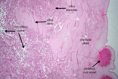
Still at low power, one may identify the chorionic plate and umbilical blood vessels, as well as the main stem of a placental villus. Like the trunk of a tree, this main stem villus will give rise to countless smaller branches which are bathed by the maternal blood of the intervillous space.
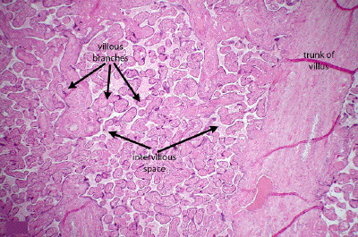
A medium power view through a portion of the placenta shows one of the main trunks of a placental villus and many other branches of varying size. The huge surface area that results from this villous branching provides adequate exchange of nutrients and waste products between fetal tissues and the maternal blood which, in life, would bathe the intervillous spaces.
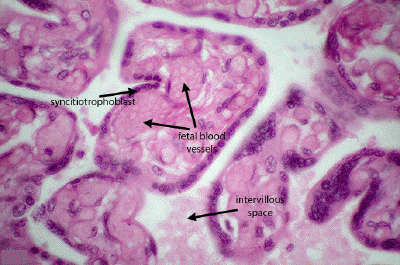
A high power view of the smallest branches of the placental villi shows the very thin placental barrier that exists in a full-term placenta. At this stage of development, only the syncitiotrophoblast separates the maternal blood of the intervillous space from the fetal blood vessels, which themselves are in close apposition to the syncitiotrophoblast.
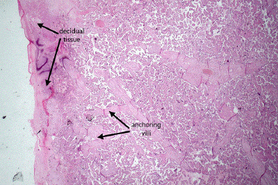
This is a low power view through the maternal side of the placenta. Some decidual tissue is seen on the left, and some of the main stems of the anchoring villi are seen approaching the maternal tissue to which they will attach.
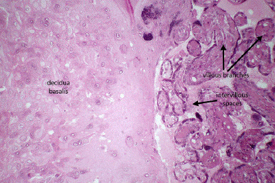
A medium power view through the maternal side of the placenta shows numerous villous branches, the intervillous spaces, and