Slide #DMS 184 [Vagina, Human, H&E]. Note the noncornified stratified squamous epithelium of the vagina and vaginal portion of the cervix ("ectocervix"). Find the well developed cervical mucous glands and the abrupt change of stratified squamous to simple columnar epithelium near the external os of the cervical canal, the junction between endocervix and ectocervix. This region of epithelial transition is clinically important since it is here that many carcinomas of the cervix develop.

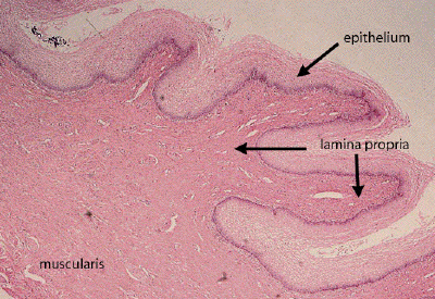
This is a low power view of a section through vagina. The vaginal mucosa is thrown up into numerous folds, or rugae, which consist of a stratified squamous epithelium supported by a well-vascularized lamina propria. A muscularis layer, comprised of smooth muscle tissue, and an outer adventitial layer, both poorly preserved in this specimen, form the wall of the vagina.

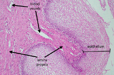
This is a medium power view of the vaginal mucosa. The cells comprising the non-keratinized, stratified squamous epithelium often have an empty appearance because of the abundant glycogen in their cytoplasm. The lamina propria is distinguished by the abundance of thin-walled blood vessels traveling through it.