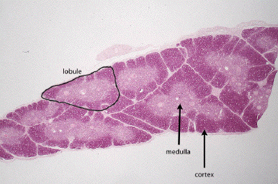
Slide #DMS 117 [Thymus, human infant]. Begin your study of the lymphoid/immune system with one of the primary lymphoid organs. (What is the other primary organ?) Use the diagram below or those in your textbook or atlas for orientation.
This slide of human infant thymus is a typical H&E stained section. Define cortex and medulla and observe that the thymus is partially divided into lobules by connective tissue septa derived from the gland capsule. Study the cortex. Note that most of the cells (thymocytes) look like small lymphocytes (type?) and are packed rather tightly together with a few interspersed large cells with large pale staining nuclei. The latter are the reticular cells of the thymus. How do these cells differ from the reticular cells found in other lymphoid organs? Examine the medulla. Note that overall there are fewer cells in this zone compared to cortex. The lymphocytes here are mostly immunocompetent but naive (virgin) T cells that have passed the selection process. How will they leave the thymus? The medulla is more heterogeneous in appearance, containing in addition to lymphocytes many macrophages, "reticular cells", abundant blood vessels and usually some thymic (Hassall's) corpuscles. The latter are balls of squamoid epithelial cells that are usually cornified and often contain keratohyaline granules. The probably are formed from defunct reticular cells but their function is still unknown.
Observe the overall histological architecture of an infant thymus in this low power view, noting the subdivision of the gland into lobules, and the discrete outer cortex and inner medulla apparent within each lobule.

This low power image of a portion of a thymic lobule shows well its division into an outer cortical zone characterized by a high density of developing T-lymphocytes (and associated support cells), and the inner medullary zone where the cells are not so tightly packed (and where the resident cells have greater amounts of cytoplasm so the dark staining nuclei are farther apart). Within the medulla, one may find Hassall's corpuscles, the rather pleomorphic structures that are unique to thymic tissue.
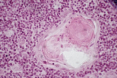
At higher magnification, observe the architecture of a Hassall's corpuscle. These structures, of unknown purpose, often show some concentric arrangement (and sometimes keratinization) of the thymic reticular cells from which they are derived.
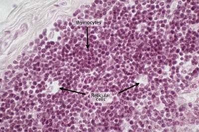
Returning to the thymic cortex, note the abundance of developing T-cells or thymocytes. Their scant cytoplasm allows for close packing of these cells at this stage of their development. Amidst these 'small dark blue cells' notice the much larger, epithelial-derived, thymic reticular cells characterized by much more abundant cytoplasm (here appearing extracted).