Slide #DMS 118 [Thymus, Monkey, PTS]. This is a 1.5 µm thin section stained by a technique similar to H&E. It is from a more mature animal, so the cortex is diminished and thymic corpuscles are more abundant in the medulla. Otherwise, look for the same features as described above. Because this is a thinner section and tissue is better preserved, more cellular detail is visible. Look for the peculiar capillaries in the cortex contributing to the blood-thymic barrier.
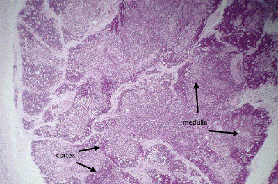
This is a low power image of a section through the thymus, probably from a somewhat older animal than that seen in the previous set of images. However, the lobular architecture of the gland, and cortical and medullary zones are still definable.
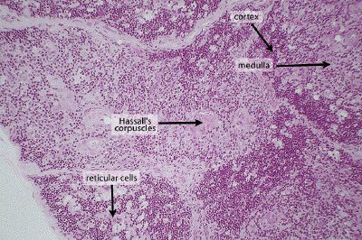
At higher power, note the dense clusters of thymocytes characterizing the cortex of the thymic lobule. Numerous large, pale thymic reticular cells are interspersed among the thymocytes, an appearance described by some as a 'starry sky' pattern. In the more extensive, pale-staining medullary region, one may define Hassall's corpuscles, seen to better effect in the next image.

In this high power image at the border between cortex and medulla, note the density of thymocytes typical of the cortex and the occasional thymic reticular cell found amongst them. A small Hassall's corpuscle is seen in the medulla.
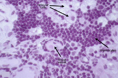
In addition to developing thymocytes and associated reticular cells, the thymus is also home to the blood-thymus barrier. These small blood vessels have a typical endothelial wall that is, in turn, ensheathed by both macrophages and thymic reticular cells that may regulate the movement of material from the blood space into the thymic parenchyma.