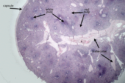
Slide #DMS 122 [Spleen, Human, H&E]. First, identify the capsule and trabeculae. Note that the largest trabeculae contain both arteries and veins whereas those of intermediate size contain only veins. Scattered smooth muscles cells may be seen within the substance of the trabeculae. A reticular cell stroma provides support for the central regions of the spleen. Next, identify the areas of red and white pulp. Note that the white pulp is arranged so as to surround small (=central) arteries thus forming a periarterial lymphatic sheath (PALS). The PALS is mainly populated by T cells. In places lymphoid nodules with germinal centers may develop in the PALS, in which case the central artery appears eccentrically-located. As in lymph nodes, these nodules are the focus of B cells. In the intervening red pulp you will see cellular strands, the splenic cords (of Billroth) separating large, thin-walled sinusoids. You should be able to trace the flow of blood through the spleen from trabecular arteries to trabecular veins. What is the significance of the "closed" vs. "open"circulation debate?
In this low power view of the spleen, appreciate that this is another of the encapsulated lymphoid organs, but rather than its parenchyma being arranged into a defined cortex and medulla, the spleen exhibits aggregates of lymphocytes known as the white pulp surrounded by a well-vascularized, less densely cellular red pulp. Connective tissue trabecula run throughout the spleen and provide a highway for blood vessels to enter and exit the splenic tissue.
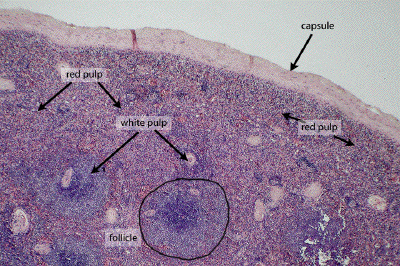
At somewhat higher power, the connective tissue capsule of the spleen is evident, as are the two parenchymal components of the spleen, the red pulp and the white pulp. The red pulp consists largely of the splenic sinusoids and a loose aggregation of several different cells types. The white pulp represents dense aggregates of lymphocytes which may be organized into either lymphoid follicles, such as those found in other lymphoid organs, or peri-arteriolar lymphatic sheaths, seen to better effect in subsequent images.
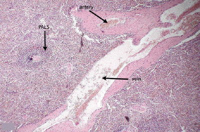
This rather low power image of the spleen show a large connective tissue trabecula through which is running a trabecular vein and a trabecular artery. In the midst of the red pulp, one can see a periarteriolar lymphatic sheath (PALS), one form of the white pulp of the spleen.
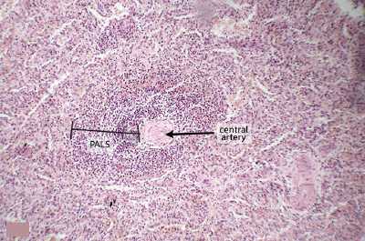
At higher power, one can define the central artery (arteriole) running through the core of the peri-arteriolar lymphatic sheath (PALS). The PALS itself consists largely of a dense aggregation of T-lymphocytes.
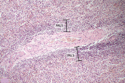
This medium power image shows a longitudinal cut through a central artery and its surrounding peri-arteriolar lymphatic sheath (PALS). As will be seen in a later image, a lymphoid follicle (B-cell dependent) is often found in close association with a PALS (T-cell dependent). The highly vascular red pulp is seen peripheral to the white pulp of the PALS.
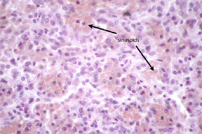
At high power, one can appreciate the typical architecture of the red pulp. Large splenic sinusoids (filled with red blood cells) are surrounded by a mixture of lymphocytes, macrophages, plasma cells, and reticular cells, most of which are best identified with specific labeling techniques. More details of the red pulp architecture are seen in the subsequent preparation.