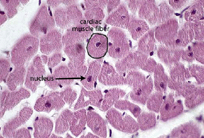
Slide #DMS 082 [Heart, Purkinki fibers, H&E]. Find areas where the branching of cardiac muscle fibers is evident; establish that they form a three-dimensional network. Note the position of the nucleus and the arrangement of myofibrils within individual muscle fibers.
Note in longitudinal section (limited areas of the slide) intercalated discs; striated patterns; central nuclei (dark blue; below); inter-bundle connective tissue; and blood vessels.

A high power view through a cross section of cardiac muscle fibers demonstrates well the centrally-located nucleus that helps to distinguish this tissue from skeletal muscle. The myofibrillar architecture of the muscle cells is also seen to good effect in this image.