Slide DMS091 [Ox spinal cord smear; toluidine blue and eosin]. Study the multipolar motor neurons when prepared by smearing fresh spinal cord gray matter between two coverglasses, drying the smear, and then staining with toluidine blue and eosin. This preparation is intended primarily to show the proximal portions of the dendrites and axons of these multipolar nerve cells as well as the Nissl substance. In most instances, the axon has been broken off. The coarse Nissl material in the cell bodies is readily recognizable and may be arranged linearly because of pressure exerted on it by the comparatively invisible neurofibrils. Relate the intense basophilia of the Nissl substance to its high content of ribonucleic acid. Under the electron microscope, these areas are characterized by the presence of endoplasmic reticulum and ribosomes. Scan the slide at low powerand note the variation in the size and appearance of the neurons. Study the morphology of the dendrites and attempt to find an axon hillock (most likely you will be unsuccessful). Are any neuroglial cells visible in this slide?
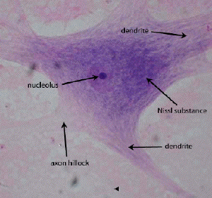
Note the nucleus with the prominent nucleolus; the rough endoplasmic reticulum (Nissl substance) that extends into the proximal dendrites; the axon hillock; and the surrounding neuropil.