Slide DMS092 is a transverse section of either human or rodent spinal cord stained by a silver impregnation technique. This technique stains dark brown to black the myelinated nerve processes (axons and dendrites) and the cytoplasmic neurofibril network of larger neurons. The remainder of the tissue stains faint yellow to pinkish-orange.
Note that the spinal cord is roughly cylindrical in shape in cross section and is composed of an inner butterfly or H-shaped core of gray matter surrounded by an outer cortex of white matter. (In general, gray matter consists mainly of nerve cells and the proximal portions of their processes imbedded in neuroglia; white matter consists mainly of myelinated nerve fibers imbedded in neuroglia.)
Gray matter. Note the anterior and posterior columns (horns) of gray matter, united by a thin gray commissure containing the small ependymal-lined central canal. Observe the large motor neurons (nerve cells) in the anterior column. Many of these cells contain darkly stained neurofibrils. The neurons are also partially outlined by darkly stained nerve cell processes. Aside from the neurons and their processes the remainder of the gray matter substance consists of supporting neuroglia cells and numerous small blood vessels (both poorly stained). Keep in mind that the amount of gray matter at any level of the spinal cord is related to the amount of skeletal muscle innervated at that level. Thus, the amount is greatest within the cervical and lumbosacral enlargements of the cord, which innervate the muscles of the upper and lower limbs respectively.
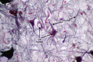
Neurites: At high power, the intricate meshwork of neurites (axons and dendrites) entangling the neuron cell bodies (perikarya) may be fully appreciated.
White matter. Note the division of spinal cord white matter into posterior, lateral and anterior columns. As in other regions of the CNS, the white matter consists here of a mixture of nerve fibers (myelinated and unmyelinated), neuroglial cells, and blood vessels. Nerve cells are usually absent from white matter. Most of the nerve fibers are myelinated which accounts for the white color of white matter (in fresh preparations) and the intense dark staining in fixed, silver-impregnated sections.