Slide DMS026 [Kidney, rabbit, x.s., PAS+H]. The PAS technique stains the basement membrane of all epithelial lined structures. Note that the patterns of the basement membranes demonstrate the architecture of the kidney quite well. The glomerular capillaries are clearly demonstrated. The denser accumulations of PAS+ material within the glomeruli are mesangial matrix. The prominent basement membrane of the tubules demonstrates their orientation. It is important to note that the epithelial cells are usually capable of regeneration so long as their basement membrane is intact. Note that the cortex contains mostly proximal convoluted tubules which can be identified by their PAS positive brush border.
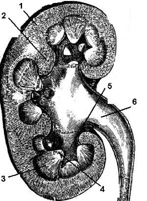
Human Kidney: 1. Cortex; 2. Renal columns; 3. Medulla; 4. Papilla of medulla; 5. Calyx; 6. Renal pelvis.