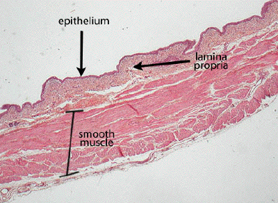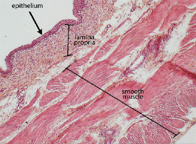
Slide #DMS 031 [Urinary bladder; H&E]. There is a typical urothelium and a lamina propria (dense inner and less dense outer layers), no submucosa and a thick outer muscular layer (smooth muscle) in which bundles of fibers go in all directions. (Some textbooks say three layers of muscle cells can be identified).

This is a low power view of a section through the bladder. The lining epithelium, supporting lamina propria, and smooth muscle wall are all seen here, and at higher power in the next image.

At this higher power, again note the transitional epithelium that lines the lumen of the bladder, the supportive, fibroelastic connective tissue of the lamina propria, and the substantial bundles of smooth muscle forming the bulk of the wall. The relative thickness of the various, rather ill-defined layers of the smooth muscle of the bladder wall will vary according to where the section was actually taken.

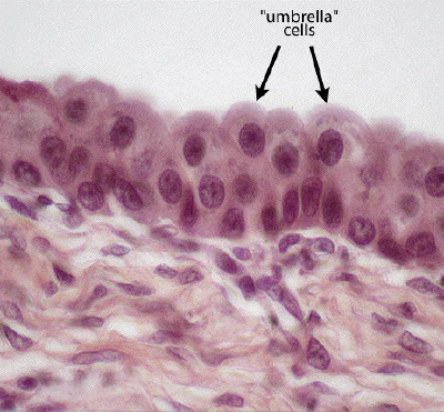
A high power view of the transitional epithelium of the bladder reveals, once again, the classic morphology of this type of epithelium, with the apical-most layer of cells assuming an 'umbrella' or pillow-like morphology when fixed in the relaxed (non-distended) condition.