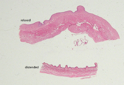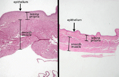
Slide #DMS 042 [Urinary bladder; relaxed, distended]. There are two sections: one of a bladder fixed in its distended state and one fixed empty. The changes in the thickness of the epithelium are striking. What features of the epithelial cells accommodate for stretching? The bladder has a lamina propria (dense inner and less dense outer layers), no submucosa and a thick outer muscular layer (smooth muscle) in which bundles of fibers go in all directions. (Some textbooks say three layers of muscle cells can be identified).
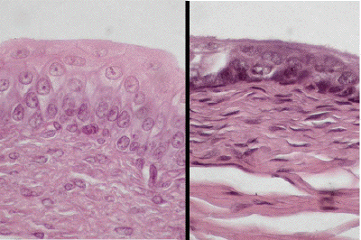
Look for an area with typical transitional epithelium, with puffy, dome-shaped surface cells. Sometimes these cells are binucleated.
The connective tissue of the lamina propria is quite dense. Because the deeper portion of this coat is a somewhat looser connective tissue, some people refer to the deeper layer as submucosa. There is no muscularis mucosae to make two separate layers, however.
The muscularis externa is thick. Although some bundles of smooth muscle run spirally, the general plan is for three layers of muscle: inner and outer longitudinal, and middle circular.
The outermost coat is an adventitia for most of the surface of the bladder, since the organ is retroperitoneal in position. Notice blood vessels and nerves running in all connective tissue layers.

This slide contains two sections of bladder, one fixed while in a relaxed state and the other fixed when the bladder was distended.

At low power, with both images taken at the same magnification, appreciate the significant thinning of the wall of the bladder in its distended state (image on right) compared to the empty relaxed bladder on the left. The lamina propria separates the transitional epithelium from the smooth muscle wall of the bladder which includes three often ill-defined layers of smooth muscle.

At high power, with both images taken at the same magnification, one may compare the differences in thickness and cell morphology of the transitional epithelium lining the relaxed (left) versus the distended (right) bladder. Lamina propria is seen beneath the epithelial layer.